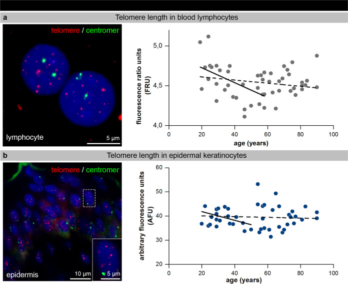
Fig. 3. Telomere-length heterogeneity in blood lymphocytes and epidermal keratinocytes.
a IF micrographs of telomere (hybridized with telomere-specific PNA probe, red) and centromere 2 (green) FISH staining in human blood lymphocytes (left panel). Graphic presentation of relative telomere-length measurements in blood lymphocytes of donors of varying age (n = 51) are given as fluorescence ratio units (FRU). Solid trend line indicates age-dependent relative telomere-length reduction up to 60 years. b IF micrographs of telomere (red) and centromere 2 (green) staining in human epidermal keratinocytes (left panel) of tissue sections. Graphic presentation of the mean relative telomere length as arbitrary fluorescence units per nucleus (AFU) in epidermal keratinocytes of donors of varying age (n = 51). Solid trend line indicates an age-dependent telomere-length reduction up to 50 years.

