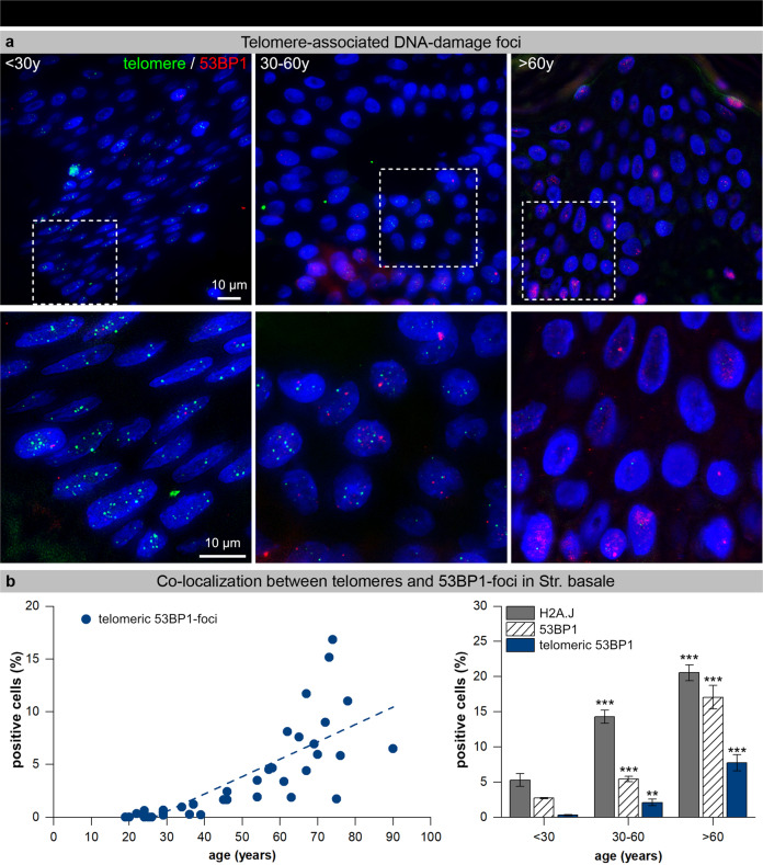
Fig. 4. Telomere-associated DNA damage foci in epidermal keratinocytes.
a Micrographs of telomere FISH signals (green) and 53BP1 IF signals (red) in human epidermal keratinocytes of young (<30 y), middle-aged (30–60 y) and aged (>60 y) skin. Selected areas are shown below at higher magnification. b Graphic presentation of epidermal keratinocytes quantification with telomeric 53BP1-foci in human skin of donors of varying age (left panel, n = 53). Graphic presentation of epidermal keratinocytes quantification with H2A.J expression, 53BP1-foci, and telomeric 53BP1-foci in the stratum basale of donors of varying age. Data are presented as mean±SE; **p < 0.01, ***p < 0.001 significant statistical difference to younger age-group.

