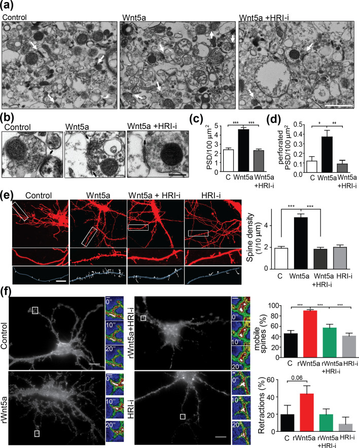
Figure 7.
HRI kinase mediates Wnt5a-dependent increase in dendritic spine density. EM images of synaptosomal fractions from mouse brain. Representative images showing the number of PSDs (a,c) and the number of multiple synaptic contacts (b,d) after the treatment with control or Wnt5a conditioned medium or co-incubation withWnt5a and HRI-i. Quantifications of the synaptic contacts and number of multiple synaptic contacts per 100 µM were included. Scale bars: 1 μm and 200 nm, respectively. Bars show the mean ± SEM. N = 3 independent experiments. Control (50); Wnt5a (59); Wnt5a+ HRI-i (55) photos (*p 0.05, **p = 0.0008, ***p = 0.0001) (e) EGFP-transfected mouse hippocampal neurons (DIV10) and IMARIS 3D reconstruction. Representative images of cropped neurites from hippocampal neurons treated with control or Wnt5a conditioned medium, incubated in the presence or absence of the HRI inhibitor. Quantification of the number of spines per 10 μm of neurite. Bars show the mean ± SEM. N = 5 independent experiments. Control (82); Wnt5a (129); Wnt5a+ HRI-i (86); HRI-I (105) dendrites (***p = 0.0001). (f) EGFP-LifeAct-transfected hippocampal neurons in the absence (control) or presence of rWnt5a, rWnt5a+HRI-i or HRI-i alone for 1 h. Insets show the fluorescence intensity of one representative protrusion measured during the indicated time (shown in each inset, 0, 10, 20 s). The graphs show the percentage (%) of mobile protrusions and the % of retraction movement for each condition. N=3 independent experiments. Control (7); rWnt5a (12); rWnt5a+HRI-I (7); HRI-I (5), number of cells from 3 independent experiments. (*p 0.05, **p < 0.001, (***p = 0.0001).

