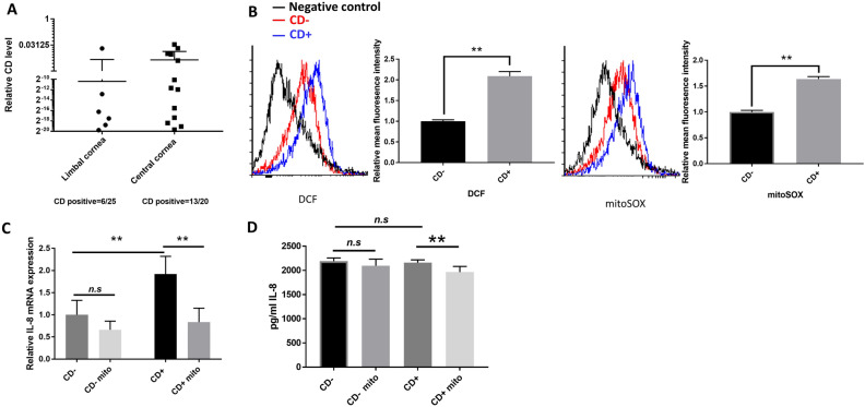
Figure 1.
Oxidative stress in CD+ keratocytes up-regulates the IL-8 expression. (A) CD/total mtDNA ratios in 45 human subjects quantified by multiplex-qPCR. 26 values were zero and are not included on the graph. (B) Flow cytometric detection of DCFH-DA and mitoSOX signal in CD+/− keratocytes (n = 3). (C) mRNA expression of IL-8 in CD+/− keratocytes. Cells either treated with 10 μM mitoTEMPO (Mito), or not, for 24 h (n = 3). (D) Quantification of secreted IL-8 level using ELISA. Cells treated, or not treated, with 10 μM mitoTEMPO for 24 h (n = 3). Values are means ± SD. n.s. (not significant); *P < 0.05, **P < 0.01.

