Figure 3.
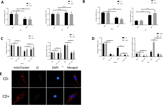
Higher level of mtdsRNA in the cytoplasm of CD+ keratocytes compared to in that of CD− keratocytes. (A) The relative level of mtDNA in the pellet fraction (PF) or cytosolic fraction (CF) measured by multiplex qPCR (n = 3). (B) Relative mtRNA in whole-cell lysate (WCL) and CF of CD+/− keratocytes measured by multiplex qPCR (n = 3). (C) Relative mtRNA in CF of CD+ keratocytes 24 h after 10 μM mitoTEMPO treatment (n = 3). (D) Mitochondrial RNA level with/without Terminator 5′-phosphate-dependent exonuclease treatment (TE) in WCL and CF (n = 3). (E) Representative fluorescence image of dsRNA in CD+/− cells. mitoTracker (red), J2 foci (greed) and DAPI (blue) were merged in the right column (merged). Values are means ± SD. n.s. (not significant); *P < 0.05, **P < 0.01.

