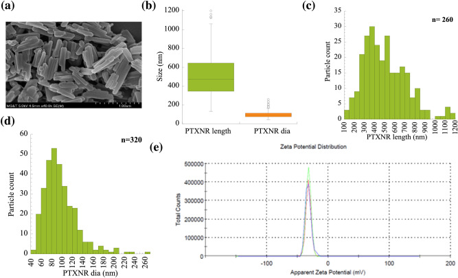
Figure 1.
Characterization of PTXNRs: (a) Scanning electron microscopy (SEM) image of PTXNRs. (b) Particle size: Percentile plots of the PTXNR-TTZ particle length and diameter were analyzed using ImageJ/ Fiji software. (c) Size distribution of PTXNR particle length. (d) Size distribution of PTXNR particle diameter as calculated analyzing SEM images. The number ‘n’ denotes the particle population count for the analysis; and (e) Zeta potential (ξ) of PTXNR is -32.6 ± 4.8 mV in water. Three colors indicate three different experiments.

