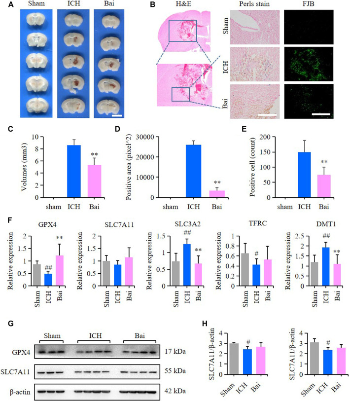
FIGURE 8.
Effect of baicalin on brain injury in ICH model mice. (A) Representative pictures of the hemorrhagic lesion in the mice of different groups. Bar = 0.5cm. (B) Representative images of Prussian blue staining and FJB staining of brain slices in the mice of different groups. Bar = 200μm. (C) Hematoma volume quantitative data of different groups (n = 6) (D) Relative statistics of Prussian blue labeling density in different groups (n = 4). (E) Relative fluorescence intensity statistics of FJB staining in different groups (n = 4). (F) The expression of GPX4, SLC7A11, SLC3A2, TFRC and SLC11A2 (DMT1) in the perihematoma brain tissues of mice were detected by RT-qPCR (n = 6) (G) The expression of GPX4 and SLC7A11 in the perihematoma brain tissues of ICH model mice were detected by western blot (H) Quantification data of western blot in different groups (n = 4). Experimental values were expressed as means ± SD. # p < 0.05 and ## p < 0.01 compared with the sham operation group and **p < 0.01 compared with the ICH group.

