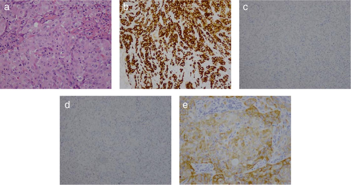
FIGURE 2.

Immunohistological characteristics of case 2, Immunohistochemical analysis of lung cancer tissue. (a) Hematoxylin and eosin staining (200×); (b) ALK ventana (DF53, 200×), (c) negative staining for TTF‐1 (200×), (d) Naspin‐A (200×); (e) positive staining for CK56 (200×)
