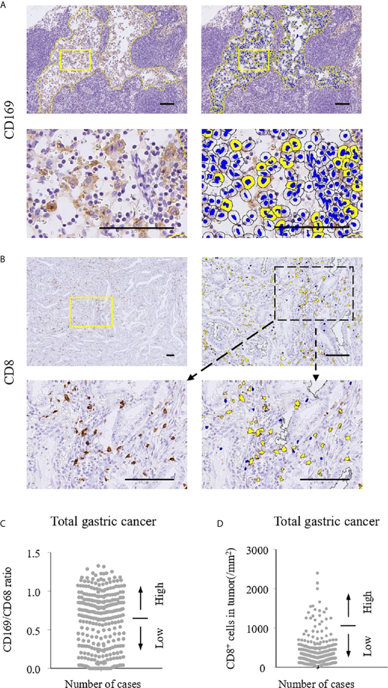
Figure 1.

Immunohistochemistry and cell counting system. The numbers of CD169+ and CD68+ cells in regional lymph nodes (RLNs) and CD8+ cells in tumors were evaluated using HALO 2.3 as described in Materials and Methods. Scale bar = 100 μm. Representative immunohistochemistry (IHC) stains of CD169 (A) and CD8 (B) are shown. Positive cells in selected areas surrounded by yellow lines were counted automatically by HALO 2.3. (C) Number of CD169/CD68 ratio in lymph node sinus macrophages (LySMs). (D) Number of CD8 expressions in primary tumor. Patients were divided into two groups according to their CD169/CD68 ratio: < 0.65 was defined as low and ≥ 0.65 was defined as high. With regard to CD8+ cells, the patients were divided into two groups according to cell density: < 287/mm2 was defined as low and ≥ 287/mm2 was defined as high.
