Figure 2.
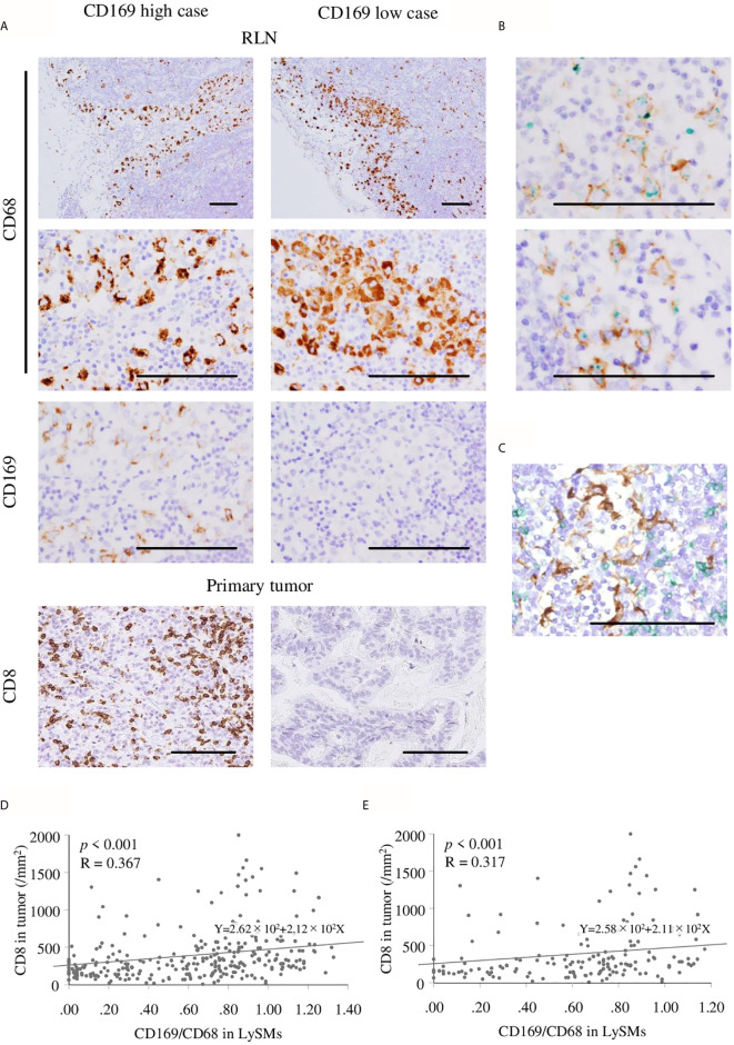
Immunohistochemistry of CD169+ and CD68+ macrophage in regional lymph nodes (RLN), and CD8+ cells in primary tumor. Scale bar = 100 μm. (A) Representative figures of immunohistochemistry (IHC) images from CD169 high and low cases are shown. Lymph node sinus macrophages (LySMs) were positive for CD68 in both two patients, although, CD169 expression differed. High infiltration of CD8+ T cells in primary tumor tissues was seen in a CD169high case and low infiltration of CD8+ T cells in primary tumor tissues was seen in a CD169low case. (B) Double IHC of CD68(green) and CD169(brown) showed CD169 was expressed on CD68-positive macrophages. Correlation between the number of CD8+ T cells in primary tumor tissues and CD169/CD68 ratio in LySMs were tested by Spearman’s correlation test. (C) Double IHC of CD8(green) and CD169(brown) showed the direct cell-cell interaction between LySM and T cells in sinus area. Scatter plots of total (D) and in advanced (E) gastric cancer cases were shown. RLN, regional lymph node.

