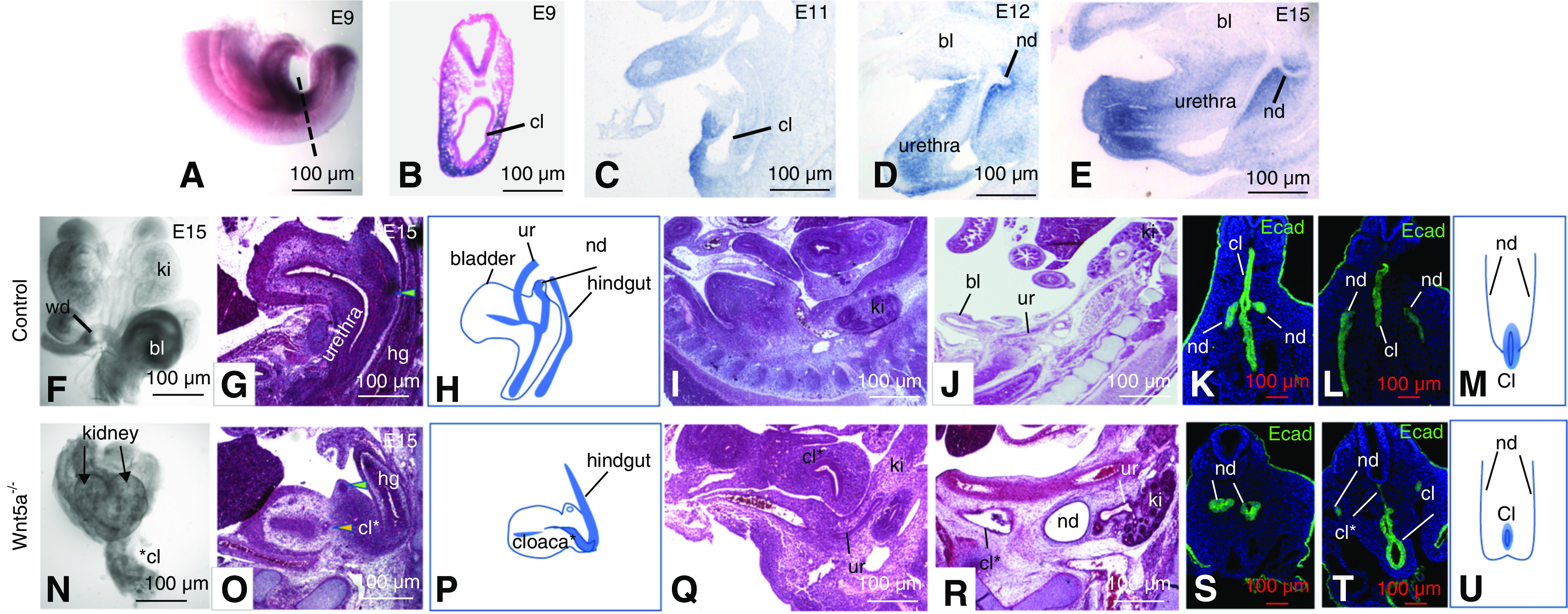
Figure 3.

Wnt5a expression and its role in lower urinary tract morphogenesis. (A–E) In situ hybridization analysis. (A) Wnt5a expression at E9 in a whole mount embryo. (B) Section through a whole mount embryo showing Wnt5a expression surrounding the cloaca. (C–E) Wnt5a expression in sagittal sections from embryos at E11 (C), E12 (D), and E15 (E). (F and N) Whole-mount urogenital tracts from a control (F) and Wnt5a a mutant (N). (G and O) Hematoxylin and eosin–stained sections through the bladder/urethra and hindgut of controls (G) and Wnt5a mutants (O). Green arrowheads point to the nephric ducts. Yellow arrowhead in (O) points to the distal ureter. (H and P) Schematic representation of histology shown in (G and O). (I and Q) Sagittal sections through Wnt5a mutants at E12. (J and R) Sagittal sections through a control (J) and Wnt5a mutant embryo (R). (K, L, S, and T) Ecad staining (green) reveals nephric duct and cloacal epithelia at E9, in controls (K and L) and Wnt5a mutants (L and T). (M and U) Schematic showing nephric duct insertion into the cloaca in a wild-type embryo (M) and in a Wnt5a mutant embryo (U). bl, bladder; cl, cloaca; cl* truncated cloaca in Wnt5a mutant; ki, kidney; nd, nephric duct; ur, ureter; wd, Wolffian duct.
