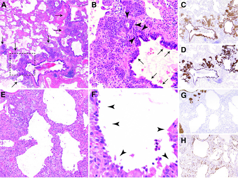
Figure 1.
Histopathology of trimethoprim–sulfamethoxazole–associated fulminant respiratory failure: diffuse alveolar injury with delayed epithelialization. (A) Low magnification (×20) shows diffuse alveolar denudation and peribronchiolar basaloid pods (PBPs, arrows), with thickened alveolar walls. (B) High-power magnification (×200) of the rectangular area in A shows an absence of pneumocytes, relative sparing of bronchioles, and prominent PBPs (previously “squamous metaplasia”), which are proliferating regenerative basaloid/squamoid cells adjacent to terminal bronchioles that are focally contiguous (arrowheads) with ciliated bronchiolar epithelium (arrows). (C and D) Pancytokeratin (C) and keratin 5/6 (D) immunohistochemical stains highlight the diffuse lack of alveolar epithelium, relatively spared bronchiolar epithelium, and basaloid nature of PBPs. (E and F) Thickened alveolar walls lack hyaline membranes or pneumocytes (E) and instead are lined by macrophages (arrowheads) (F). (G and H) Pancytokeratin stain (G) confirms the lack of alveolar pneumocytes, which are replaced by CD68-positive macrophages (H).

