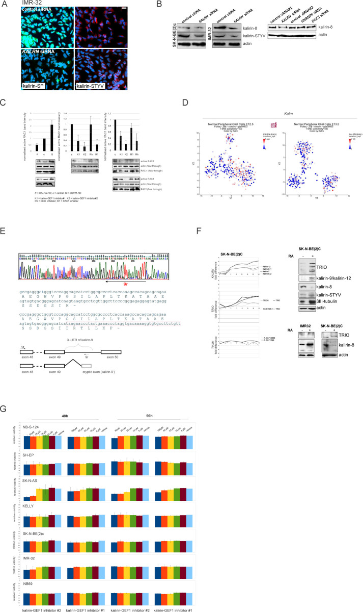
Figure S7. Modulation of KALRN levels in ADRN NB with RNAi and RA-induced differentiation.
(A) Immunolabelling of kalirin in IMR-32 after KALRN RNAi. Scale bar 20 μm. (B) Western blot analysis of kalirin-8 and kalirin-STYV levels in IMR-32 and SK-N-BE(2)c after KALRN RNAi and kalirin-8 levels in IMR32 after KALRN RNAi or additional negative control siRNAs: control siRNA #2, HNRNPK siRNA, and DISC1 siRNA. (C) RAC1 activity in IMR-32 and SK-N-BE(2)c cell treated with kalirin-GEF1 inhibitor#1 (10 μM), kalirin-GEF1 inhibitor#2 (5 μM), RHOA inhibitor (3 μM), RAC1 inhibitor (10 μM), or after SOX11 and KALRN RNAi. (D) Kalrn expression in t-SNE-resolved E12.5 and E13.5 sympathetic precursors: sympathoblasts, Schwann cell precursors, bridge population and chromaffin cells (Furlan et al, 2017). (E) Sequencing electrophoregrams showing 3′-UTR of kalirin-9 isoform (top) and the exon scheme based on the results of sequencing (bottom). Location of stop codon is marked with “-.” (F) KALRN, TRIO, and TIAM expression in SK-N-BE(2)c (left) after RA (10 μM) treatment. x-axis indicate timepoints in hours. Western Blot analysis of kalirin, TRIO, and βIII-tubulin in SK-N-BE2c after 72 h of RA-treatment (top right) and of TRIO and kalirin-8 in IMR-32 and SK-N-BE(2)c after 72 h of RA treatment (bottom right). (G) Cell viability of NB cell lines treated with vehicle, kalirin-GEF1 inhibitor#2, or kalirin-GEF1 inhibitor#1. Values are reported as mean percent ± SD of vehicle-treated control.

