Figure 3.
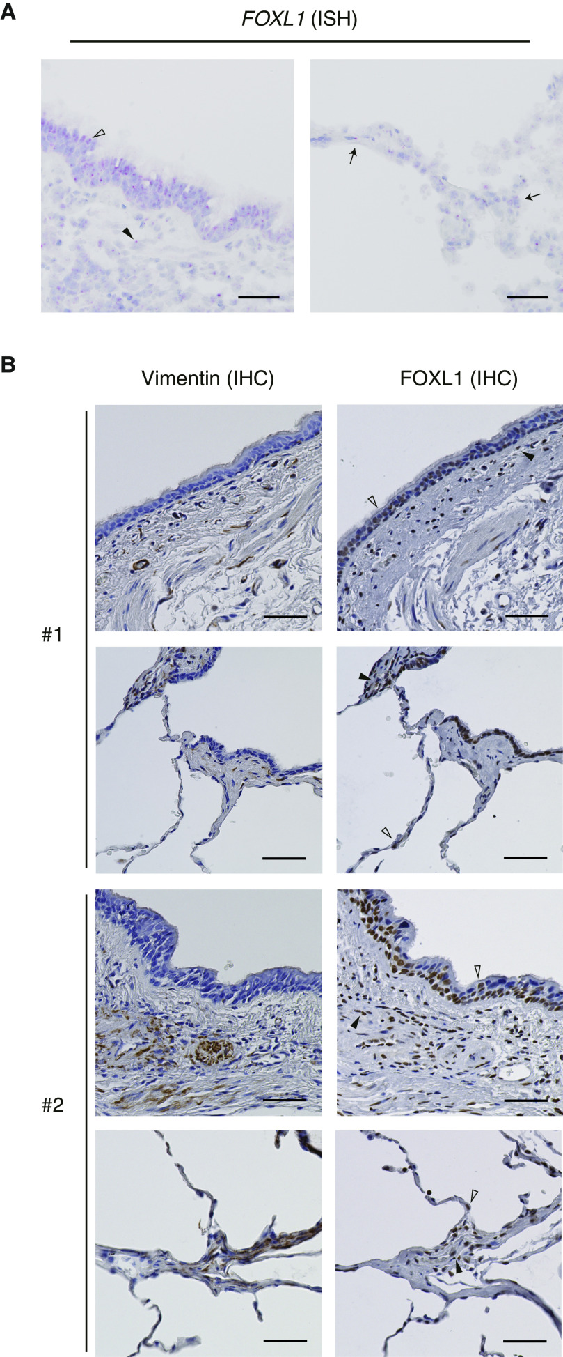
Expression of FOXL1 in human lung tissue. (A) FOXL1 expression detected by RNA ISH in normal lung tissue. Left: FOXL1 was positively stained in the nuclei of both epithelial cells (open arrowhead) and interstitial cells (solid arrowhead) in the airway. Right: Nuclear FOXL1 positivity was found in the alveolar cells (arrows). Scale bars, 50 μm. (B) Immunohistochemistry (IHC) for vimentin (left) and FOXL1 (right) in normal lung tissue. Two different subjects (#1 and #2) are presented. FOXL1 was positively stained in the nuclei of both epithelial cells (open arrowheads) and interstitial cells (solid arrowheads) in the airway (top) and the alveolus (bottom). Note that nuclear FOXL1 staining is observed in the interstitial cells positive for vimentin. Scale bars, 50 µm. ISH = in situ hybridization.

