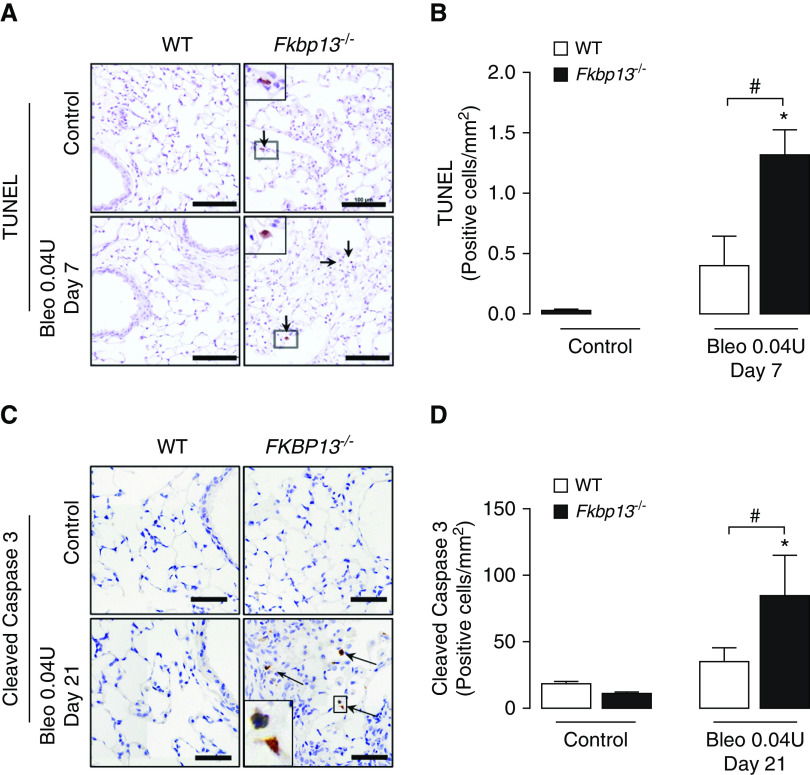
Figure 6.
FKBP13−/− lungs have increased amount of UPR activation and apoptotic cells. WT and FKBP13−/− mice (n = 6–8/group) were treated with the low dose (0.04 U/mouse) of Bleo, and lung tissues were assessed for UPR and apoptosis markers by using IHC, in situ hybridization, and Western blot. (A and B) TUNEL staining and quantification of positive cells (arrow) at Day 7 by using HALO image-analysis software. Scale bars, 100 μm. (C and D) Cleaved caspase 3 immunostaining and quantification of positive cells (arrow) at Day 21 by using HALO image-analysis software. Scale bars, 100 μm. Images were acquired using the automated Olympus VS120 slide scanner, which utilizes a stitching algorithm to reconstruct the whole specimen from overlapping image tiles. Magnified regions shown in inset image. *P < 0.05 versus untreated mice. #P < 0.05 between genotypes.

