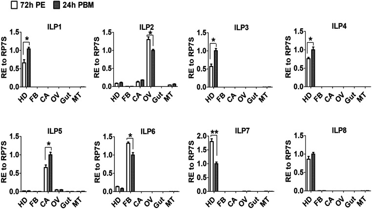
Fig. 1.
Tissue distribution of ILP transcription in female Ae. aegypti mosquitoes. Relative expression levels of ilp genes in the head (HD), isolated fat body (FB), carcass (CA, abdominal wall without the fat body), ovary (OV), gut, and Malpighian tube (MT) at 72 h PE and 24 h PBM. Relative expression levels in abundant group at 24 h PBM are represented as 1, with corresponding adjustments in other values. Data represent three biological replicates with 30 individuals in each and are shown as mean ± SEM *P < 0.05, **P < 0.01.

