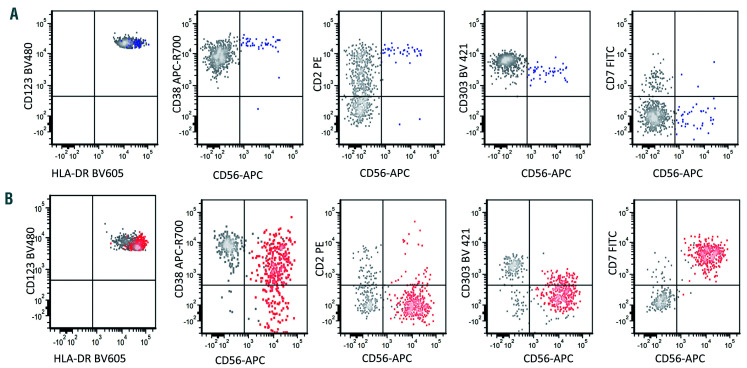
Figure 4.
Representative cases of reactive and neoplastic CD56+ plasmacytoid dendritic cells. Gray: CD56– reactive plasmacytoid dendritic cells (PDC); blue: CD56+ reactive PDC; red: CD56+ neoplastic PDC. (A) CD56+ reactive PDC are consistently positive for CD2 and CD303, negative for CD7. CD38 expression is bright. (B) In contrast, neoplastic CD56+ neoplastic PDC are often negative for CD2 with decreased to negative CD303 expression. CD7 expression is often positive and CD38 expression level is often decreased. Focusing on CD56– PDC (gray) in both panels (A) and (B), these cells are positive for CD303 and CD38. CD2 shows a bimodal pattern of expression (both negative and positive cells present). A small subset of reactive PDC is CD7+ and these CD7+ reactive PDC are negative for CD56.

