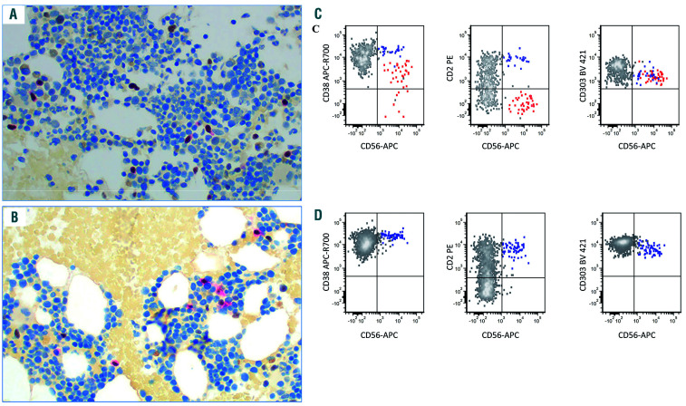
Figure 6.
Immunostain and flow cytometric analysis of a case of blastic plasmacytoid dendritic cell neoplasm before and after transplantation. (A, B) Immunostain using a dual-color TCF4/CD123 double stain showed scattered plasmacytoid dendritic cells (PDC) in both samples, before (A) and after (B) transplantation. (C, D) Flow cytometric analysis showed that a subset of PDC (red) in the pre-transplant sample (C) was aberrant (decreased CD38, negative CD2, decreased CD303 expression) whereas all PDC in the post-transplant sample (D) showed a normal immunophenotype. CD56+ reactive PDC are highlighted blue in (C) and (D).

