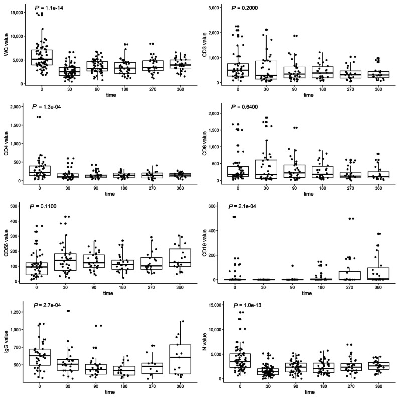
Figure 2.
Cellular and humoral immune reconstitution after axicabtagene ciloleucel (axi-cel). Box and whisker plots demonstrating cellular and immune reconstitution following treatment with axi-cel. White blood cells (WC) and neutrophils (N) were measured by complete blood counts with differential at baseline (n=85), day 30 (n=70), day 90 (n=56), day 180 (n=42), day 270 (n=32), and day 360 (n=31). CD3 T cells, CD4 T cells, CD8 T cells, CD56 natural killer cells, and CD19 B cells were measured by flow cytometry at baseline (n=58) and at day 30 (n=34), 90 (n=31), 180 (n=26), 270 (n=20) and 360 (n=19). Also shown are serum mmunoglobulin G (IgG) levels at baseline (n=58) and at day 30 (n=34), 90 (n=31), 180 (n=19), 270 (n=17) and 360 (n=17) after treatment with axi-cel. Boxes demonstrate first quartile, median and third quartile values. Whiskers show the data ranges. Dots represent individual patients. P-values are calculated by the Kruskal-Wallis test to assess significant differences in the indicated cell type after axi-cel infusion.

