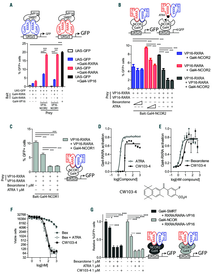
Figure 5.
Interaction of co-repressors NCOR and SMRT with natural retinoic acid receptor (RAR)A : retinoid X receptor (RXR)A heterodimers. (A) Mammalian twohybrid assay schema. 293T cells co-transfected with plasmids encoding the reporter: UAS-GFP; “bait”: Gal4-RXRA or Gal4-RARA; and “prey”: VP16-SMRT or VP16-NCOR. The percentage of GFP+ cells was assessed 48 hours (h) after transfection by flow cytometry in triplicate. A vector encoding Gal4-VP16 fusion was used as a positive control. (B) Reversely, 293T cells were co-transfected with plasmids encoding the reporter: UAS-GFP; “bait”: Gal4-SMRT; and “prey”: VP16-RARA and/or VP16-RXRA. Cells were treated with increasing concentrations of all-trans retinoic acid (ATRA) and bexarotene (0, 100 nM and 1 mM) in triplicate. The percentage of GFP+ cells was assessed 48 h after transfection by flow cytometry. (C) 293T cells were co-transfected with plasmids encoding the reporter: UAS-GFP; “bait”: Gal4-NCOR; and “prey”: VP16-RARA and/or VP16-RXRA. Cells were treated with ATRA and bexarotene (1 mM) in triplicate. The percentage of GFP+ cells was assessed 48 h after transfection by flow cytometry. (D and E) MLL-AF9 leukemia cells derived from UAS-GFP bone marrow and transduced with MSCV-Flag-Gal4-RXRA-IRESmCherry retrovirus (MLL-AF9 Gal4-RXRA cells) or MSCV-Flag-Gal4-RARA-IRES-mCherry retrovirus (MLL-AF9 Gal4-RARA cells) were treated as indicated, replated and total viable cells in 50 mL assessed after 96 total h of treatment in duplicate. (F) MLL-AF9 cells were treated as indicated, replated and total viable cells in 50 mL assessed after 96 total h of treatment in duplicate. (G) 293T cells were co-transfected with plasmids encoding the reporter: UAS-GFP; “bait”: Gal4-NCOR or Gal4- SMRT; and “prey”: VP16-RARA and/or VP16-RXRA. Cells were treated with ATRA, bexarotene, and CW103-4 as indicated in triplicate. The percentage of GFP+ cells was assessed 48 h after transfection by flow cytometry. *P<0.05, ***P<0.001, t-test with Welch’s correction.

