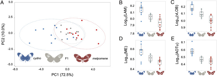
Fig. 3.
Intermediate brain morphology in H. m. rosina × H. c. chioneus F1 hybrids. (A) Variation in H. m. rosina (red), H. c. chioneus (blue), and hybrid (gray) brain morphology in a principal-component analysis of all segmented neuropils and rCBR. (B–E) Examples of neuropils with intermediate volumes in hybrids (B and C), or melpomene-like volumes (D and E) in F1 hybrids; lamina (LAM), ventral lobula (vLOB), medulla (ME), and anterior optic tubercule (AOTu).

