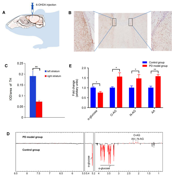
Figure 5.

The BLF metabolome under the condition of Parkinson's disease. A, Brain infection with 6‐hydroxydopamine (6‐OHDA) to selectively destroy dopaminergic and noradrenergic neurons in the right striatum. (n = 7). B,C, 21 days after the infection, TH‐positive neurons decreased sharply in the right striatum compared to the left striatum. Bars: 200 μm. D, Differential‐metabograms of BLF from control rats (n = 8) and 6‐OHDA injection rats (n = 7). Metabolites with red color are significantly changed between groups. MUDA were used to evaluate the significance of difference in metabolite concentrations between groups. E, Relative change fold of significant metabolites (acetoacetate (AA), N‐acetylglycoprotein (N‐AG), O‐acetyl‐glycoprotein (O‐AG) and α‐glucose). Student's t‐test was used to evaluate the significance of difference in metabolites between between the PD group and control group. *P < 0.05, **P < 0.01.
