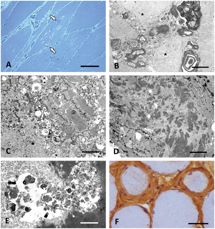
Figure 1.

Characteristic morphological findings in vacuolar myopathies. A. Prominent vacuoles containing granular osmiophilic material (arrows) in GNE myopathy (P14). Semithin section, toluidine blue. Scale bar = 40 µm. B,C. Large osmiophilic membranous and granular inclusions indicative of altered autophagy combined with granulofilamentous material and other myofibrillar breakdown products (asterisks) in desminopathy (P7). EM. Scale bar in B = 3 µm, in C = 1.5 µm. D. Filamentous bundles characteristic for ZASPopathy in P10. EM. Scale bar = 3 µm. E. Large autophagic vacuole containing pleomorphic granular and membranous material in a case of Bethlem myopathy (P11) caused by COL6A2 mutation. EM. Scale bar = 2 µm. F. Prominent endomysial fibrosis with concentric accumulation of collagen and of fibroblasts around individual muscle fibers in P11. Paraffin section, Coll VI immunohistochemistry (brown). Scale bar = 30 µm.
