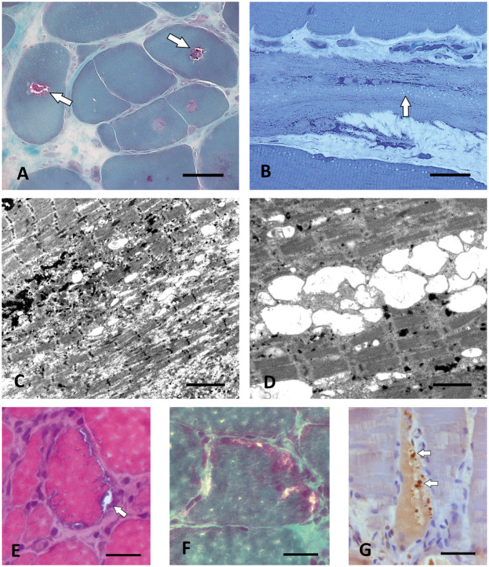
Figure 2.

Rimmed vacuoles in sarcotubular myopathy and FSHD2. A. Central rimmed vacuoles (white arrows) in P1.2 with compound heterozygous TRIM32 mutations. Cryostat section, Gömöri trichrome. Scale bar = 30 µm. B. Numerous smaller empty vacuoles arranged in rows typical for sarcotubular myopathy in P1.1, the brother of P1.2 who carried the same compound heterozygous TRIM32 mutations as P1.2. Semithin section, toluidine blue; scale bar = 30 µm. C,D. By EM, the small vacuoles in the biopsy of P1.1. At least partially correspond to dilations of the sarcoplasmic reticulum. Scale bar in C = 4 µm, in D = 2 µm. E. Muscle biopsy of the FSHD2 patient P6 showing subsarcolemmal rimmed vacuoles (arrow). Cryostat section, H&E. Scale bar = 20 µm. F. Subsarcolemmal rimmed vacuoles in the same biopsy as in (E). Cryostat section, Gömöri trichrome. Scale bar = 20 µm. G. The autophagic vacuoles in P6 contain pTDP‐43‐immunoreactive granular material (arrows). Paraffin section. Scale bar = 30 µm.
