Figure 4 1.

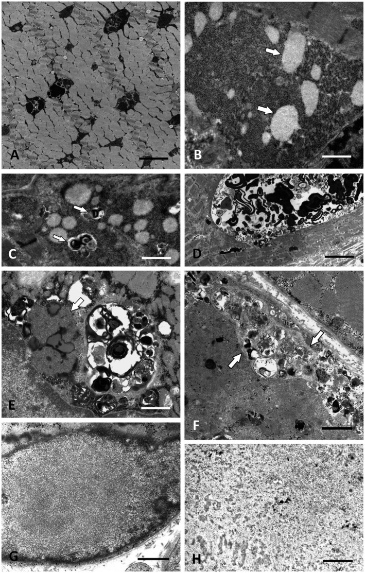
Ultrastructure of GSD II, GSD XV, chloroquine myopathy and cases without identifiable gene defect. A. Intermyofibrillar deposits of excess glycogen in GSD II (Pompe disease; P12). Scale bar = 3 µm. B. Muscle biopsy of P13 with novel compound heterozygous GYG1 (GSD XV) mutations showing excess glycogen with distinct round clusters of less osmiophilic fine granules, possibly representing partially degraded glycogen (arrows). Scale bar = 2 µm. C. P13: Autophagic vacuoles (arrows) associated with the glycogen deposits. Scale bar = 2.5 µm. D. P13: Large intermyofibrillar autophagic vacuole. Scale bar = 8 µm. E. Autophagic vacuoles combined with extensive accumulations of lipofuscin and curvilinear material (arrow) typical for chloroquine myopathy (P16). Scale bar = 2 µm. F. Arrows marking the membranous borders of exocytosed material between the plasma membrane and the basal lamina in P21. However, no mutation in LAMP‐2 or any other myopathy gene was found in this patient by WES. Scale bar = 2 µm. G. Distinct OPMD‐like tubulofilaments within a myonucleus in P27, who did not harbor a detectable PABPN1 repeat expansion or any other mutation in a myopathy gene. Scale bar = 0.5 µm. H. Deposition of granulofilamentous material in P19 suggested the diagnosis of a desminopathy/myofibrillar myopathy, but no defect in any of the relevant known genes was detected by WES. Scale bar = 0.5 µm.
