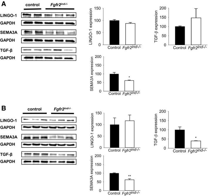
FIGURE 7.

Remyelination inhibitor expression in the acute and chronic phase of EAE. Representative western blots and quantification are shown for the acute and chronic phase of EAE. (A) In the acute phase of EAE there were no differences in remyelination inhibitor LINGO‐1 and SEMA3A expression between Fgfr2ind −/− mice and controls. Interestingly, remyelination inhibitor TGF‐β expression was less in Fgfr2ind −/− mice. (B) In the chronic phase Fgfr2ind −/− mice showed less expression of remyelination inhibitors SEMA3A and TGF‐β. LINGO‐1 expression was not regulated in the chronic phase of EAE. n = 2–3 in acute phase and n = 3 in chronic phase, data are presented as mean ±SEM. *p < 0.05; **p < 0.005.
