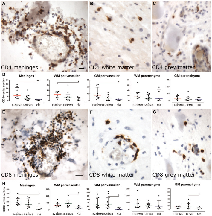
Figure 3.

T cell infiltrates in the SPMS spinal cord. Immunohistochemical detection of CD4+ T cells in different compartments of the spinal cord of SPMS cases (A–C). The number of CD4+ T cells was greater in the meninges and in white and grey matter perivascular cuffs in F+ SPMS compared to controls (D). CD8+ T cells were occasionally seen as modest infiltrates in the meninges and at higher numbers than CD4+ T cells in perivascular spaces and parenchyma of the spinal cord white and grey matter (E–G). There was a large and variable extent of CD8+ infiltrates of the cord meninges, which differed between F+ SPMS and controls but not between F+ and F− SPMS (H). Scatter dot plot of total immunopositive cells per section, from three sections per case, with median and interquartile range indicated. Kruskal–Wallis and Dunn's multiple comparison posttest. *P < 0.05. Scale bar: 40 µm. Ctrl, control; F+ SPMS, lymphoid‐like structure positive SPMS; F− SPMS, lymphoid‐like structure negative SPMS.
