Figure 1.

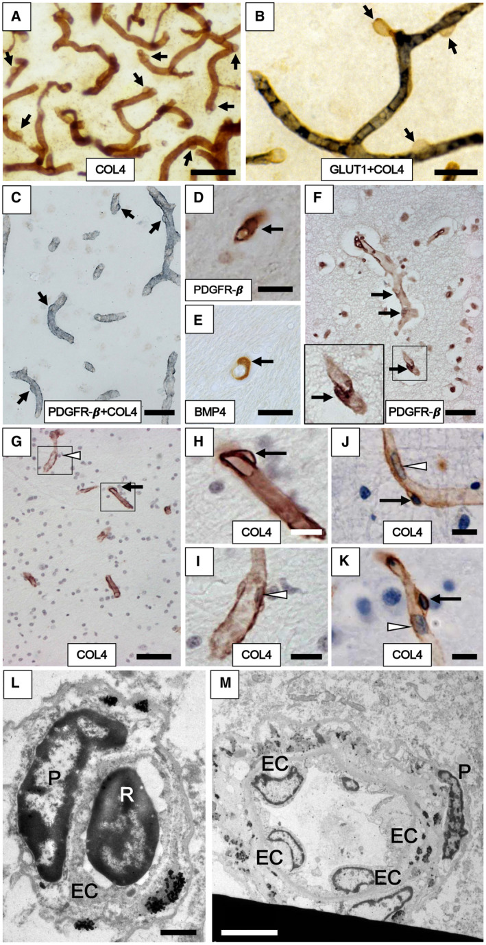
Cerebral capillaries with pericytes in the frontal cortex and WM of aging subjects. A–F. Pericytes evident as “bumps” or “crescent” shaped cells in the capillary network immunostained with antibodies to COL4 (A, G, H, I, J and K), GLUT1 (blue‐black) and COL4 (brown) (B), PDGFR‐β (brown) and COL4 (blue/gray) (C), PDGFR‐β (D, F) and BMP4 (E). Arrows show location of the pericyte cell bodies along the capillaries. G–K. Pericyte soma (black arrows in G, H, J and K) and endothelial cell (white arrowhead in G and I) and other types of cells including erythrocytes observed within the lumen (white arrowhead in J and K) in the frontal WM capillaries of the Baboon (G–I) and human (J and K). L,M. Transmission electron microscope images show pericyte cell body (P) on a WM capillary (L) and precapillary/small arteriole (M) in cross‐section from 65‐year‐old male subject. The marked differential localization between the pericyte and endothelial cell (EC) and the lumen with an erythrocyte (R for red blood cell) can be noted. L–M also assure assessment of non‐capillary pericytes or EC or smooth muscle cell nuclei were not included in the pericyte counts. Images in A and B were from 20 µm thick sections, whereas those from C to I were from 10 µm sections. Length density of frontal cortical capillaries (A) was greater than WM (B). Magnification bars: A, G = 50 µm; C, F = 30 µm; B, D, E = 20 µm; H, I, J, K = 10 µm; L = 1 µm; M = 5 µm.
