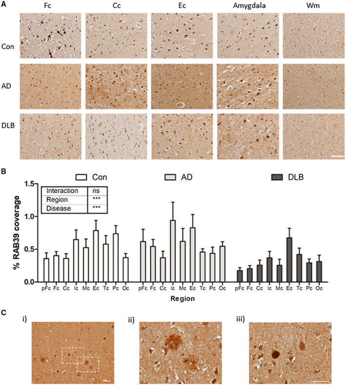
Figure 1.

Cortical distribution of RAB39B in the human brain. A. Representative 100x images of frontal (Fc), cingulate (Cc) and entorhinal (Ec) cortex from control (Con), Alzheimer’s disease (AD) and dementia with Lewy bodies (DLB) cases. Additional images from the amygdala and frontal white matter (Wm) are also shown. In cortical regions, robust somatic cytoplasmic staining was apparent (arrows) and in some cells RAB39B appeared within intracellular cytoplasmic bodies (arrowhead). B. Comparative quantification in Con, AD and DLB cases of the percentage area coverage of RAB39B immunoreactivity in the pre‐frontal (pFc), frontal (Fc), cingulate (Cc), insular (Ic), motor (Mc), entorhinal (Ec), temporal (Tc), parietal (Pc) and occipital (Oc) cortices. Data shown as mean ± SEM and output from two‐way ANOVA is stated, ***P < 0.001 and ****P < 0.0001. C. Examples of RAB39B positive neuropathological aggregates in the cortex of one individual DLB case. At 100x magnification, both Aβ plaques (magnified at 400X in ii) and Lewy bodies (LBs; magnified at 400x in iii) were evident. Scale bar in A and C i = 50 µm and in C iii = 100 µm
