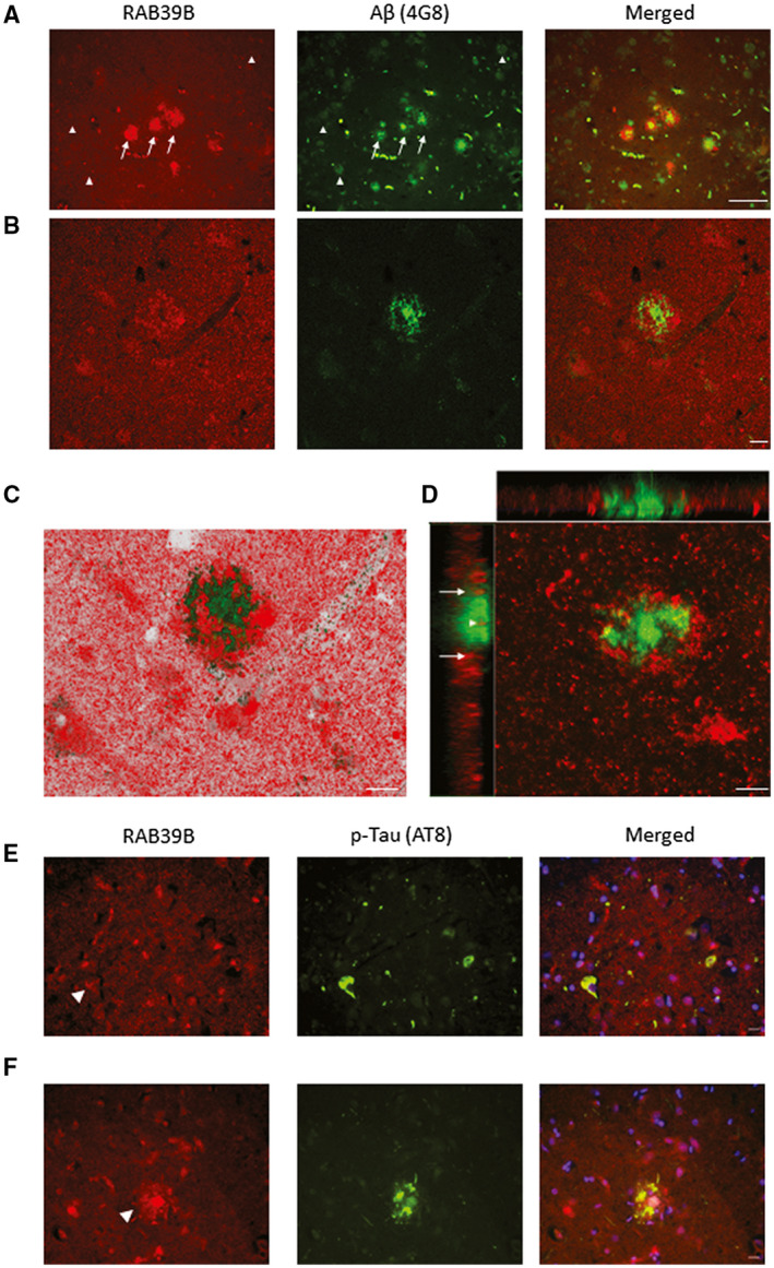
Figure 2.

Co‐aggregation of RAB39B in Aβ plaques. Representative images of frontal cortex tissue stained for RAB39B (red) and Aβ (green) in a case of AD (A‐D). A. Wide‐field 200x fluorescence micrographs demonstrated the frequent co‐localization of RAB39B within cortical dense core Aβ plaques (arrows) in contrast to diffuse Aβ deposits (arrow heads) which were RAB39B negative. Co‐aggregation of RAB39B within dense core plaques was confirmed via confocal micrograph images (B‐D). Specifically, Z‐stack analysis (D) determined RAB39B sequestration within the core of the plaque (arrowhead) in addition to the accumulation of RAB39B at the periphery of the plaque (arrows). E and F. 400x micrographs of frontal cortex tissue stained for RAB39B (red) and AT8 positive phosphorylated tau (p‐Tau; green) are presented. E. No overt accumulation of RAB39B was observed in neurofibrillary tangle bearing neurons (arrowhead). F. Co‐localization of AT8 p‐Tau within RAB39B positive plaques was observed, indicative of neuritic subtype. Slides were coverslipped with DAPI containing mounting media (blue; E‐F). Scale bar in A = 50 µm and in B‐F = 10 µm
