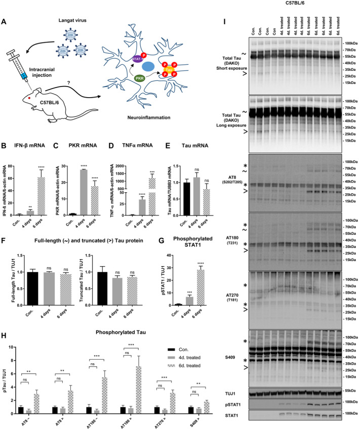
Figure 7.

Brain infection with Langat virus induces PKR upregulation, abnormal tau phosphorylations and tau truncation in C57BL/6J mice. A. Schematic figure describing experimental setup and hypothesis. In brief, C57BL/6J mice were intracranial injected with 100 focus forming units (FFU) of Langat virus for four‐ or six d. PBS injected mice served as control. Langat virus brain infection induces high viral RNA levels and interferon response associated with PKR activation. The hypothesized PKR activation on STAT1 and tau phosphorylation was assessed. B–E. mRNA levels in RNA extracted from brain homogenates were quantified by qPCR. IFN‐β mRNA (B), PKR mRNA (C) and TNF‐α mRNA (D) were normalized to β‐actin mRNA while neuronal Tau mRNA (E) was normalized to neuron‐specific class III beta‐tubulin (TUBB3) mRNA. The values in the figure exhibit the ratio between PBS injected mice and 4 or 6 days Langat virus injected mice (n (PBS) = 4, n (4 days Langat virus) = 4 and n (6 days Langat virus) = 5, **P < 0.01, ****<0.0001 student t‐test). F–H. Quantifications of the effect of 4 or 6 days Langat virus brain infection on total tau/TUJ1 (F), phospho‐STAT1/TUJ1 (G) or full‐length (~) and truncated (>) phospho‐tau epitopes/TUJ1 levels (H) (n (PBS) = 4, n (4 days Langat virus) = 4 and n (6 days Langat virus) = 5, **P < 0.01, ***<0.001, ****<0.0001 student t‐test). I. Immunoblot of total brain homogenate from C57BL/6J mice intracranial injected with PBS or 100 FFU of Langat virus for 4‐ or 6 days. using total‐ [anti‐tau (A0024, DAKO)] and pTau‐specific antibodies as well as anti‐STAT1 and anti‐pSTAT1. pSTAT1 was used as positive control for PKR activation in the brain. Sign ~ indicates size of total‐ and abnormally phosphorylated full‐length tau, > indicates size of total‐ or abnormally phosphorylated truncated tau species, while * indicates size of non‐tau bands recognized by anti‐mouse secondary antibody.
