Figure 2.

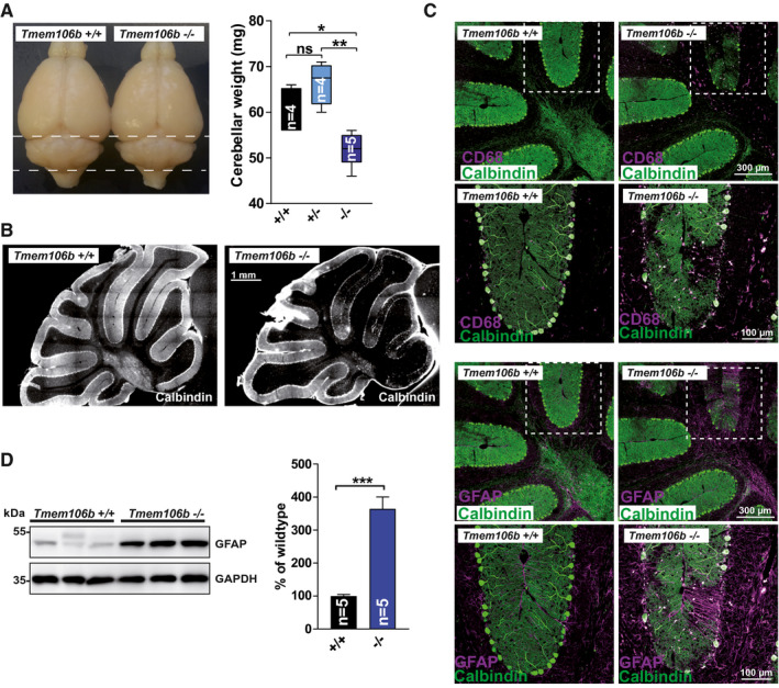
Cerebellar degeneration in aged Tmem106b KO mice. (A) Photos of representative brains of 18‐month‐old WT and Tmem106b KO mice. Cerebellar wet‐weight of WT, Tmem106b HET and Tmem106b KO mice is depicted (age: 18 months). (mean ± SEM, n = 4–5; *P < 0.05, **P < 0.01). (B) Representative immunofluorescence staining of cerebellar sections for the Purkinje cell marker calbindin‐D28‐K of WT and Tmem106b KO mice (age: 18 months). (C) Triple Co‐immunofluorescence staining of cerebellar sections from WT and Tmem106b KO mice for calbindin‐D28‐K (green) and the microglia/macrophage marker CD68 (magenta) (upper panel) and calbindin‐D28‐K (green) and the astrocyte‐marker GFAP (magenta), (lower panel). (D) Immunoblot of cerebellar extracts from WT and Tmem106b KO mice with an antibody against GFAP. GAPDH is depicted as a loading control. Quantification of the GFAP, normalized to GAPDH is shown. The mean of the values for the WT animals was set as 100%. (mean ± SEM, n = 4–5 ***P < 0.001).
