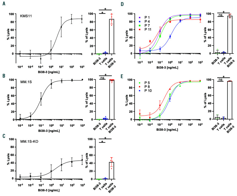
Figure 1.
Bi38-3 induces CD38-dependent T-cell-mediated lysis of multiple myeloma cells in vitro. (A-C) KMS11-luc (A), MM.1S-luc (B) and CD38-deficient MM.1S-luc (MM.1S-KO-luc) (C) multiple myeloma cell lines were co-cultured with T cells, isolated from peripheral blood samples from healthy donors, at an effector: target (E:T) cell ratio of 5:1 with increasing concentrations of Bi38-3 for 24 h. Curves represent target cell lysis, monitored by luciferase activity and expressed as the percentage of the untreated condition (0% lysis). Data are the means of independent experiments with four (A), nine (B) and four (C) different donors. Standard deviations (SD) are shown for each concentration. Histograms on the left show target cell lysis induced by Bi38-3 alone, donor T cells alone and T cells with Bi38-3 (101 ng/mL) on KMS11-luc (A), MM.1S-luc (B) and MM1.S-KO-luc (C). (D and E) Fresh tumor plasma cells were collected from buffy coat of bone marrow aspirates from myeloma patients, then CD138+ cells were purified and co-cultured with autologous CD3+ T cells isolated from peripheral blood mononuclear cells at an E:T cell ratio of 5:1 for 24 h. Cultures were analyzed by fluorescence-activated cell sorting (FACS) to monitor the number of CD138+ cells falling into the live gate. The average percentages of lysis of CD138+ cells (relative to the untreated condition) in four different patients at diagnosis (D) and three different patients at relapse (E) are shown. The error bars indicate the SD. Histograms show the average effects of Bi38-3 alone, T cells alone and T cells with Bi38-3 (102 ng/mL) on tumor plasma cells from the same four patients at diagnosis (D) and three patients at relapse (E). SD are shown and P values were determined by a two-sided Mann–Whitney U-test (*P<0.05; **P<0.01; ***P<0.001).

