
Figure 3.
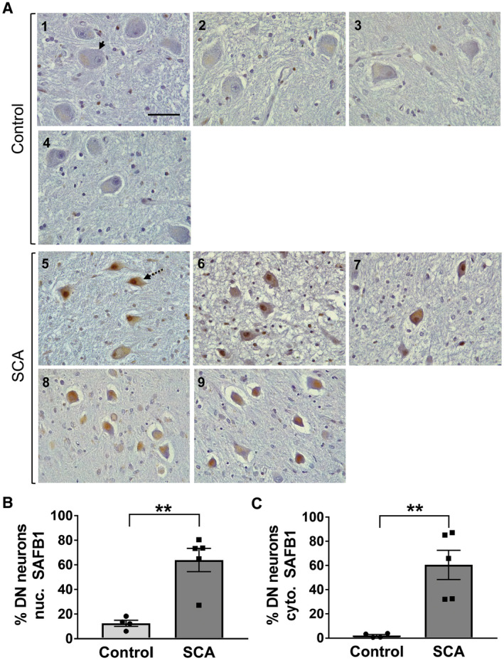
SAFB1 expression is increased in the nucleus and cytoplasm of cerebellar dentate nucleus neurons in SCA patients. A. Representative images of cerebellar dentate nucleus immunostained for SAFB1 show large principle neurons (identifiable by their morphology) within the neuropil of the dentate nucleus in control and SCA cases (arrow). Cells within SCA patient dentate nucleus appear smaller and shrunken in shape (dotted arrow) and show increased nuclear and cytoplasmic SAFB1 expression. Numbering represents patient case number, scale bar is 50 µm. B‐C. SAFB1 Immunoreactivity is expressed as the percentage of large principle neurons which are immuno‐positive for SAFB1 in either the nucleus or the cytoplasm. In SCA patients, a significantly higher percentage of neurons are positive for SAFB1 within the nuclei compared to control cases (unpaired, two‐tailed t‐test t(7) = 4.675, P = 0.0023) (B). Cytoplasmic SAFB1 is also expressed in a significantly higher percentage of neurons of SCA patients compared to control cases (unpaired, two‐tailed t‐test t(7) = 4.253, P = 0.0038) (C). **P < 0.01, ***P < 0.001. Data are presented as mean ± SEM.