
Figure 4.
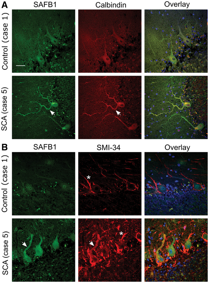
SAFB1 colocalizes with calbindin and SMI‐34 in the cytoplasm of SCA Purkinje cells. A. Calbindin immunopositivity specifies Purkinje cell soma and dendrites. SAFB1 staining is absent from the cytoplasm of control Purkinje cells but is colocalized with calbindin in SCA cases (arrows). Cell nuclei were stained using DAPI (blue). B. SMI‐34 staining, a marker of Purkinje cell damage colocalizes with SAFB1 in the soma and dendrites of SCA cerebellum (arrows) but not control tissue. SMI‐34 also stains basket cell projections (asterisk), which appear hypertrophied in SCA tissue compared to control. Scale bar is 50 µm.