
Figure 1.
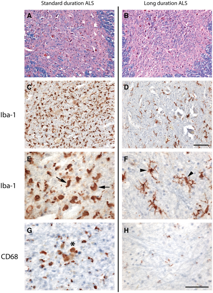
Decreased microglial cell density and resting‐type morphology within the spinal cord in long‐duration ALS. (A) and (B) Luxol fast blue/hematoxylin and eosin staining demonstrates severe degeneration and loss of motor neurons in both standard (A) and long (B) duration ALS. (C–F) Immunostaining for Iba‐1 shows increased numbers of Iba‐1‐positive microglia in standard (C) compared to long (D) duration ALS. The morphology of the microglia is altered toward a reactive phenotype with a larger, ameboid shape in standard duration ALS (E, arrows) compared to the resting phenotype of a small cell body with ramified processes in long‐duration ALS (F, arrowheads). (G and H) Foci of CD68‐positive macrophages are more frequent in standard (G, asterisk) than in long‐duration ALS (H). Scale bar in A–D, 100 μm; E‐H, 50 μm.