Figure 2.

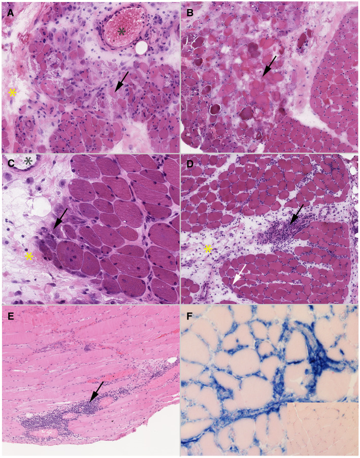
Morphological analysis. Optical microscopy. A to D: frozen sections, hematoxylin‐eosin (HE), magnification ×100. C: paraffin‐embedded sections, hematoxylin‐eosin, magnification ×100. D: frozen section, histoenzymology, alkaline phosphatase (ALP) reaction using SK‐5300 Vector Blue kit revealing ALP activity in blue, with eosin counterstaining, magnification ×200. A: case without perifascicular atrophy but with several necrotic fibers (arrow), which is not specific‐DM features. A perimysial edema is observed too (yellow star). Moreover, blood vessels in perimysium are dilated (grey star). B: muscle biopsy showing a microinfarct, frequently seen in DM. A perimysial edema is observed too (yellow star). C: case with a marked perifascicular atrophy (arrow), which is seen in only 12% of the cases, associated with the internalization of the nucleus. A perimysial edema is observed too (yellow star) as well as enlargement of blood vessels in perimysium (grey star). D: there is a mild perifascicular atrophy (white arrow) and a strong perifascicular inflammatory infiltrate is present (block arrow), which both are specific‐DM features. Finally, a perimysial edema is observed on HE stain (yellow star). E: perifascicular inflammatory infiltrate of mononucleated cells (black arrow). F: perimysial edema positive with ALP reaction (blue staining) compared to normal muscle (inset). ALP activity appears in blue, and the normal muscle in pink with the eosin counterstaining
