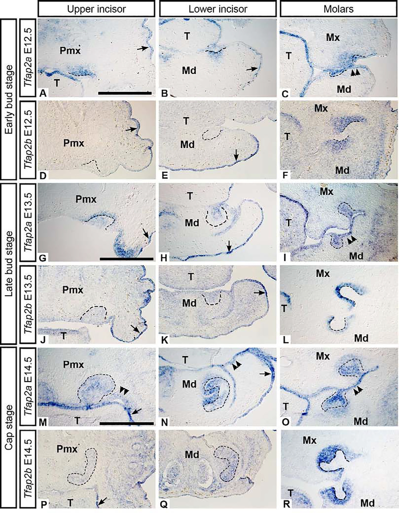
Figure 1. Bud stage (E12.5 and 13.5) and cap stage (E14.5) mRNA expression of Tfap2a and Tfap2b in wild-type mouse embryos.
Images showing mRNA transcripts detected by in situ hybridization on frontal cryosections through the upper incisor (left column), lower incisor (middle column), and molars (right column). The dental epithelium is outlined. There is minimal expression of Tfap2b in the bud stage upper and lower incisors (D-E, J-K) compared to the molar buds (F, L). Both Tfap2a and Tfap2b were detected in the surface epithelium (arrows) but only Tfap2a was present in the oral epithelium (M, N double arrowheads). Scale bars in A, G, M: 500um, all images are at the same scale. Pmx: premaxilla, Mx: maxilla, Md: mandible, T: tongue.

