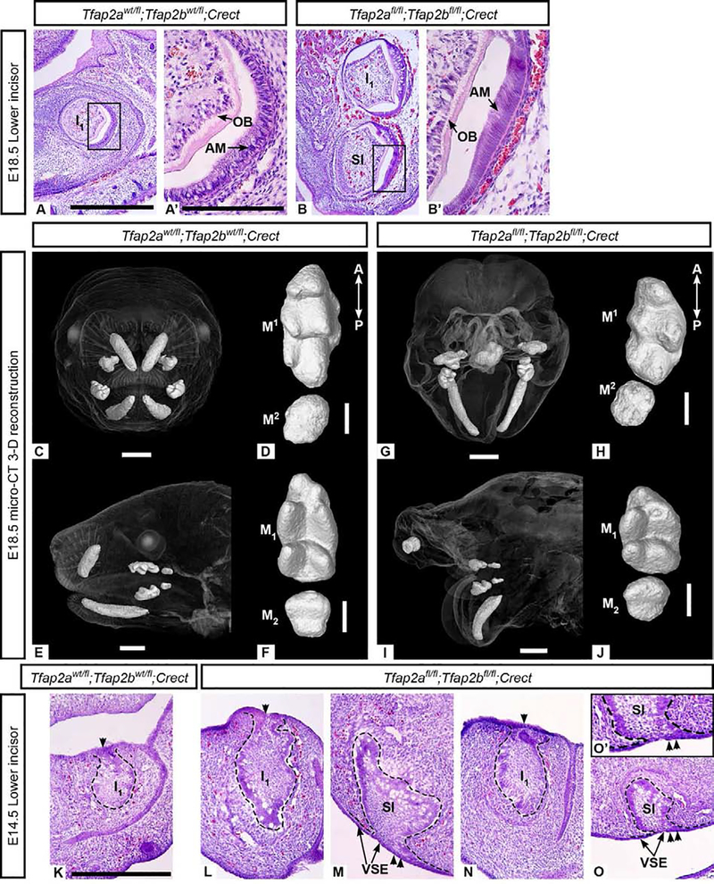
Figure 3. Tfap2afl/fl;Tfap2bfl/fl ;Crect mutant embryos have duplicated or ventrally curved lower incisors.
Hematoxylin and eosin (H&E) staining of bell stage lower incisors from control (A, A’) and epithelial mutant (B, B’) embryos revealed a supernumerary incisor ventral to I1 in the mutant mandible. This additional incisor undergoes cytodifferentiation (B’) similar to I1 in mutant (B) and control (A, A’) embryos. All mutant mandibles examined exhibited ventral curvature as seen in G and I. In the 3-D reconstructed embryo (G, I), instead of duplicated lower incisors, a single ventrally curved lower incisor was present on the right and left sides and two small right and left upper incisors were also observed. 3-D reconstructions of upper (D, H) and lower (F, J) molars show that first (M1/1) and second (M2/2) molars develop in mutants lacking epithelial Tfap2a and Tfap2b (H, J) and that the main cusps are present, though less distinct, in mutants compared with the controls (D, F). The mutant molars appear shorter along the anterior-posterior (A-P) axis than the control molars. H&E staining of frontal cryosections through cap stage lower incisors from control (K) and epithelial mutants, where I1 and a supernumerary incisor are shown in two different individuals (L-M and N-O, respectively). I1 is attached to the dorsal dental lamina (single arrowhead) in the control (K) and mutants (L, N). The supernumerary incisors are tethered to the ventral surface epithelium (double arrowheads, see the region of attachment shown in M, O, and enlarged in O’) and are positioned ventral and slightly posterior to I1. Scale bars: 500um (A), 150um (A’), 1mm (C, E, G, I), 300um (D, F, H, J, K). A and B, are to scale; A’ and B’ are to scale; K-O are to scale. AM: ameloblasts, OB: odontoblasts, SI: supernumerary incisor, VSE: ventral surface epithelium.

