Fig. 2.
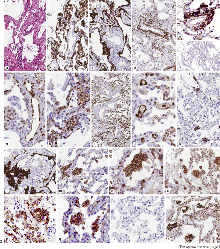
Abnormal morphology and phenotype in enlarged vascular endothelial cells: H&E (a); CK7 (b, c). Lymphocyte infiltration of vascular walls: CD3 (d), CD4 (e), and CD8 (f): perivascular lymphocytes mostly exhibit a CD3+, CD4+, CD8-negative immunophenotype. Ph-STAT3 (g): strong nuclear expression in endothelial cells. PD-L1 (h, i), and IDO (j): strong expression in capillaries and venules. CD61 (k): occasional positive megakaryocytes within interstitial capillaries. Immunohistochemical profile of aggregates of alveolar mononuclear cells: CK7 negative (l), CD11c+ (m), CD4+ (n), CD14+ (o), CD123+ (p), CD206+ (q), CD303-negative (r), and PD-L1+ (s). CK7, cytokeratin 7.

