
Figure 1: Confocal single-molecule fluorescence spectroscopy of freely diffusing molecules using Pulsed Interleaved Excitation (PIE) and four-channel detection.
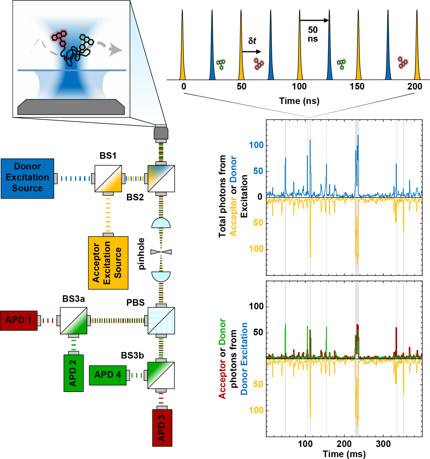
Two interleaved pulsed lasers (blue and yellow) for donor and acceptor excitation, respectively, are coaxially aligned using a dichroic mirror (BS1) and then directed into the back aperture of a high-numerical-aperture microscope objective. The spatial selection of the femtoliter confocal observation volume enables single-molecule detection at sub-nanomolar concentrations. When a single molecule diffuses through this volume, it produces a short (~1 ms) burst of donor (green) and acceptor (red) photons. The signal upon direct excitation of the acceptor is shown in yellow. Fluorescence photons are then collected by the same objective in an epifluorescence configuration, spatially separated from the excitation light using a second dichroic mirror (BS2), focused through a pinhole to reject out-of-focus fluorescence, split by polarization (PBS), and directed towards avalanche photodiodes (APDs 1–4) via a pair of dichroic mirrors (BS3a and BS3b) chosen to spectrally separate donor and acceptor fluorescence. The resulting four detection channels correspond to parallel and perpendicular polarized fluorescence from the donor and acceptor fluorophores, respectively.