Fig. 2—

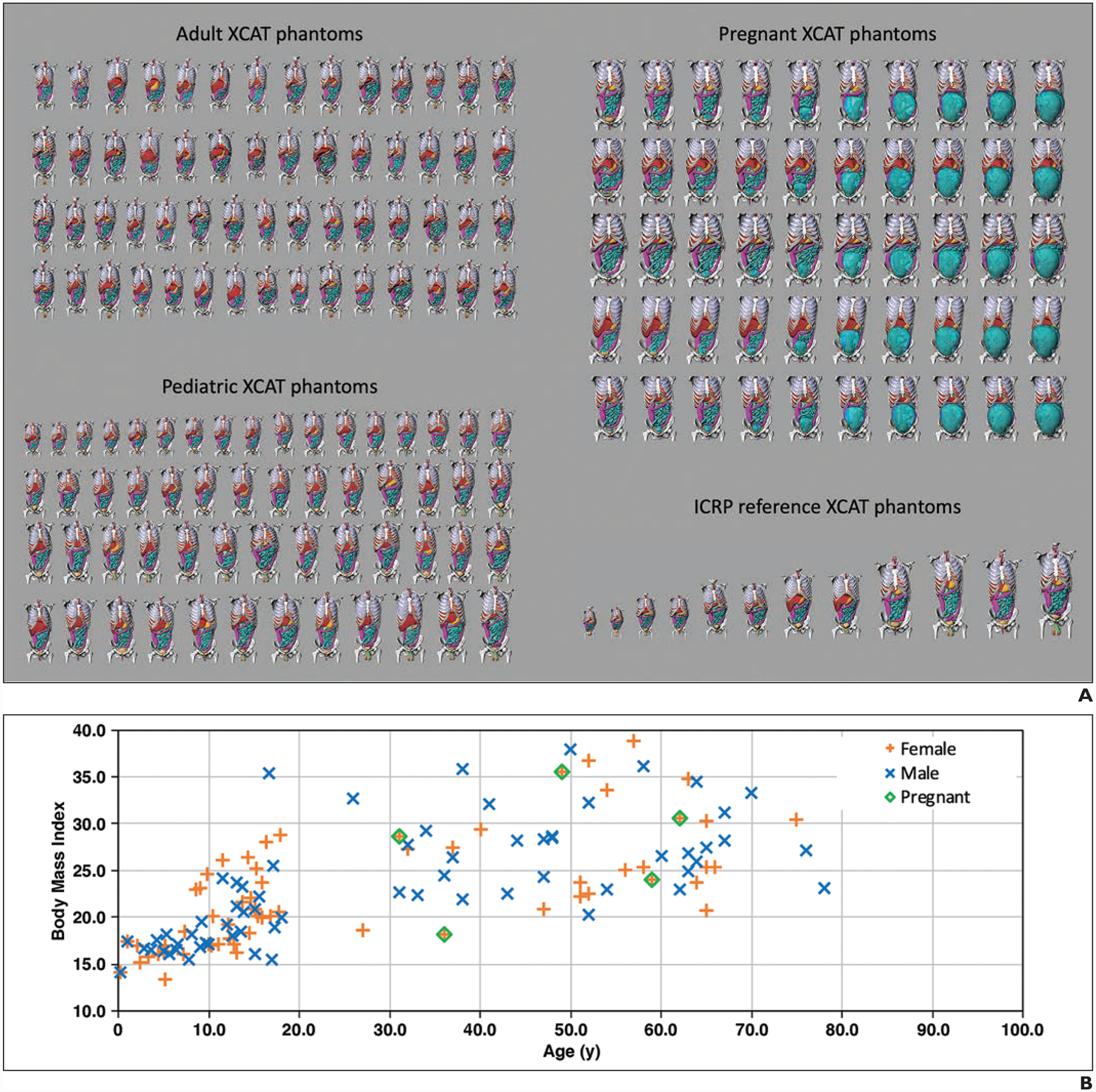
Extended cardiac-torso (XCAT) phantoms used in study.
A, Chart shows frontal views of phantoms of 58 adults (age range, 18–78 years; 23 women, 35 men), 56 pediatric patients (age range, 2–18 years; 31 girls, 25 boys), five pregnant women (gestational age range, 3–38 weeks), and 12 International Commission on Radiological Protection (ICRP) reference XCAT phantoms used in this study. Phantom skin, head, arm, and legs were removed to enhance visualization of organs in chest-abdomen-pelvis region.
B, Graph shows body mass index and age of XCAT phantom variations within population.
