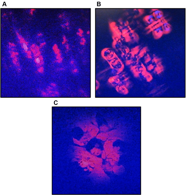
Figure 3.
Morphological changes of A549 cells after treatment with CA sponges extract, detected by dual staining of Hoechst 33342 and propidium iodide (PI). (A) The untreated cells or 0 h observation, indicated structurally normal viable cells with normal nuclei. (B) After 12 h of CA sponges extract treatment, A549 cells were expected to be apoptotic as evidenced by the cell shrinkage and the separation of condensed chromatin. (C) On 24 h observation, the plasma membrane of A549 cells had lost their integrity and formed apoptotic bodies.

