
Figure 4.
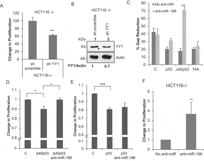
∆40p53 inhibits cell proliferation mediated by miR-186-5p
(A) BrdU cell proliferation assay performed in HCT116-/-cells transfected with sh YY1 and scrambled sh RNA as a nonspecific control (sh scramble) (n = 3). (B) Western blot analysis of cell extracts from HCT116-/- (expresses only ∆40p53) cells transfected with sh YY1 and scrambled sh RNA as a nonspecific control (sh scramble) probed with anti YY1 antibody and anti-actin antibody. (C) Wound-healing assay performed in H1299 cells co-transfected with C: GFP (vector control)/p53FL only/∆40p53 only/14A (expresses both p53FL and ∆40p53) and anti-miR-186-5p. The graph represents the percentage gap reduction in the wound n = 3). (D, E) BrdU cell proliferation assay performed in H1299 cells co-transfected with C: GFP (vector control)/p53FL only/∆40p53 only and anti-miR-186-5p. The graph represents the change in proliferation (n = 3). (F) BrdU cell proliferation assay performed in HCT116-/-cells transfected with anti-miR-186-5p.