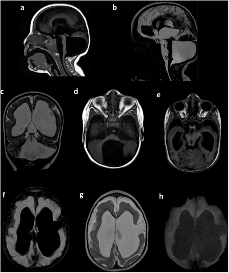
FIGURE 2.
Brain MRI. Sagittal T1-weighted (a) and sagittal constructive interference in steady state (CISS) (b) sequences showing microcephaly with a giant posterior fossa cyst, a small and displacement of the brain stem, and tightening of the posterior fossa. Coronal T2-weighted images (c) showing ventricular dilatation, cortical and white matter narrowing, and posterior fossa cyst. Axial T1-weighted images (d) showing a posterior fossa cyst. Axial T1-weighted multiplanar reconstruction (e) showing a narrow posterior fossa, cerebellar vermis with hypoplasia, and superior and lateral deviation of the cerebellum. Axial FLAIR (f) and axial T2-weighted sequences (g) showing hydrocephalus and anomalous cerebral cortex with lissencephaly and left frontal open-lip schizencephaly. Axial susceptibility-weighted image (h), with the absence of intracranial calcifications. Preliminary data of this study were previously presented in the IBRO meeting 2019 (Candelo et al., 2019).

