Figure 5.
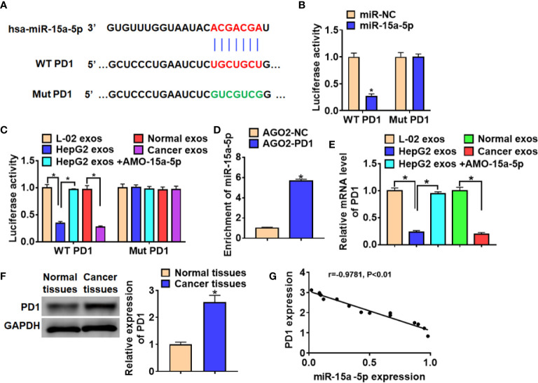
MiR-15a-5p directly targeted PD1. (A) Predicted miR-15a-5p target sequences in the 3′ UTRs of PD1 genes. (B) WT and mutant PD1 luciferase plasmids were transfected into HEK293 cells with miR-15a-5p or NC. The luciferase activity was measured by dual-luciferase reporter assay system. (C) Luciferase assay for WT and mutant PD1 activity in CD8+ T cells incubated with exosomes from L-02, HepG2, HepG2 transfected with AMO-15a-5p, healthy volunteer serum or HCC patient serum. (D) RIP assay was performed to detect the enrichment of miR-15a-5p in CD8+ T cells transfected with PD1 or NC. (E) qRT-PCR analysis for PD1 expression in CD8+ T cells incubated with exosomes from L-02, HepG2, HepG2 transfected with AMO-15a-5p, healthy volunteer serum or HCC patient serum. (F) The protein expression of PD1 in normal tissues and cancer tissues in HCC. (G) The expression correlation between of miR-15a-5p and PD1 in HCC. Data are mean ± SD; *P < 0.05.

