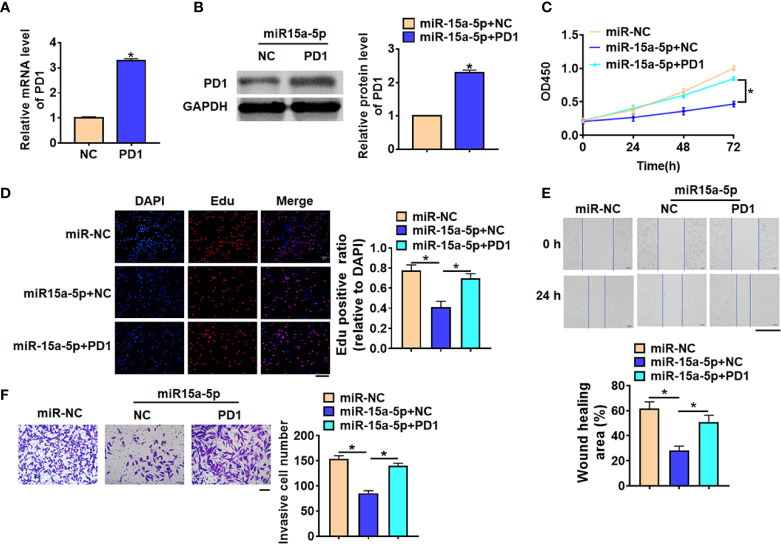
Figure 6.
Exo-miR-15a-5p suppressed HCC progression by inhibiting PD1 expression in CD8+ T cells. PD1 was forced expression in CD8+ T cells, and co-cultured CD8+ T cells with HepG2 cells transfected with miR-15a-5p. (A) The transfection efficiency of PD1 was determined by qRT-PCR. (B) The protein expression of PD1 was determined by western blot. (C) CCK8 for viability of HepG2 cells. (D) Edu assay for proliferation of HepG2 cells. Scale bar, 100 μm. (E) Wound healing assay for migration of HepG2 cells. Scale bar, 500 μm. (F) Transwell assay for invasion of HepG2 cells. Scale bar, 100 μm. Data are mean ± SD; *P < 0.05.

