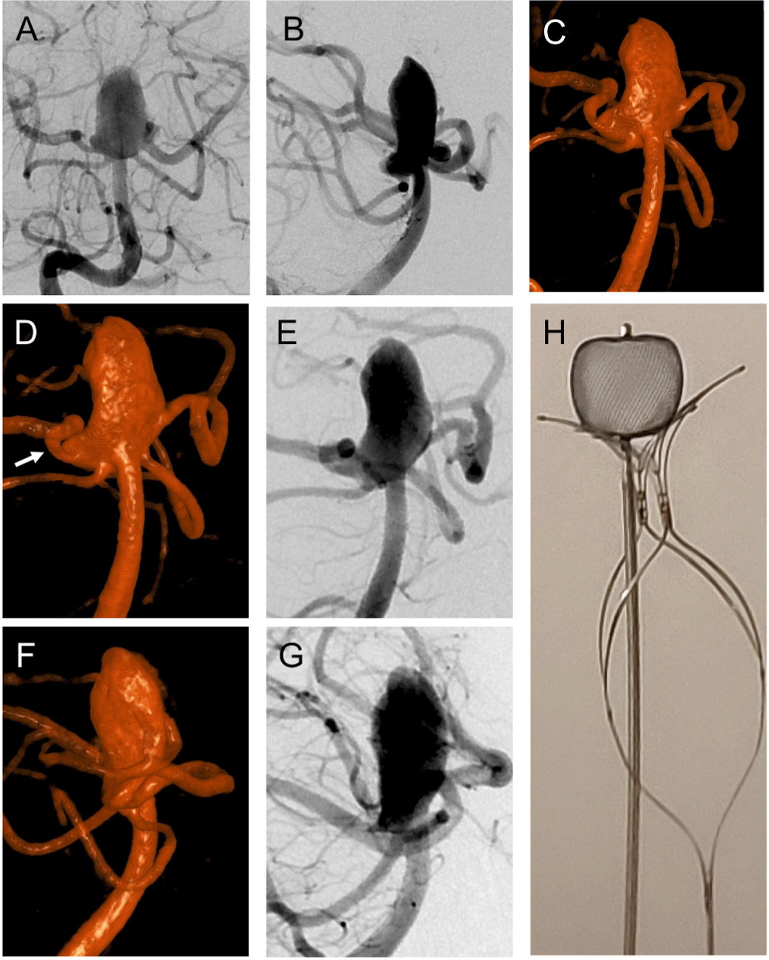
Fig. 2.

(A) Right vertebral arteriogram in frontal projection shows wide-necked, large basilar apex aneurysm incorporating both PCA origins. (B) Lateral projection of right vertebral arteriogram shows the relationship of both PCAs and SCAs to the aneurysm. (C) Volume-rendered reformat of the flat panel CTA showing the extension of the aneurysm into the right PCA origin. (D) Volume-rendered reformat of the flat panel CTA in the right anterior oblique Water’s projection for treatment. (E) Right anterior oblique Water’s projection (same as D) of right vertebral arteriogram for treatment. (F) Volume-rendered reformat of the flat panel CTA in the right posterior oblique Schuller’s projection for treatment. (G) Right posterior oblique Schuller’s projection (same as F) of right vertebral arteriogram for treatment. (H) Ex vivo demonstration of WEB SLS 11 × 9.6 mm atop PulseRider Y-shape with 10.6 mm arch width.
