


Figure 1 |. Endoscopic appearance of refractory EoE.
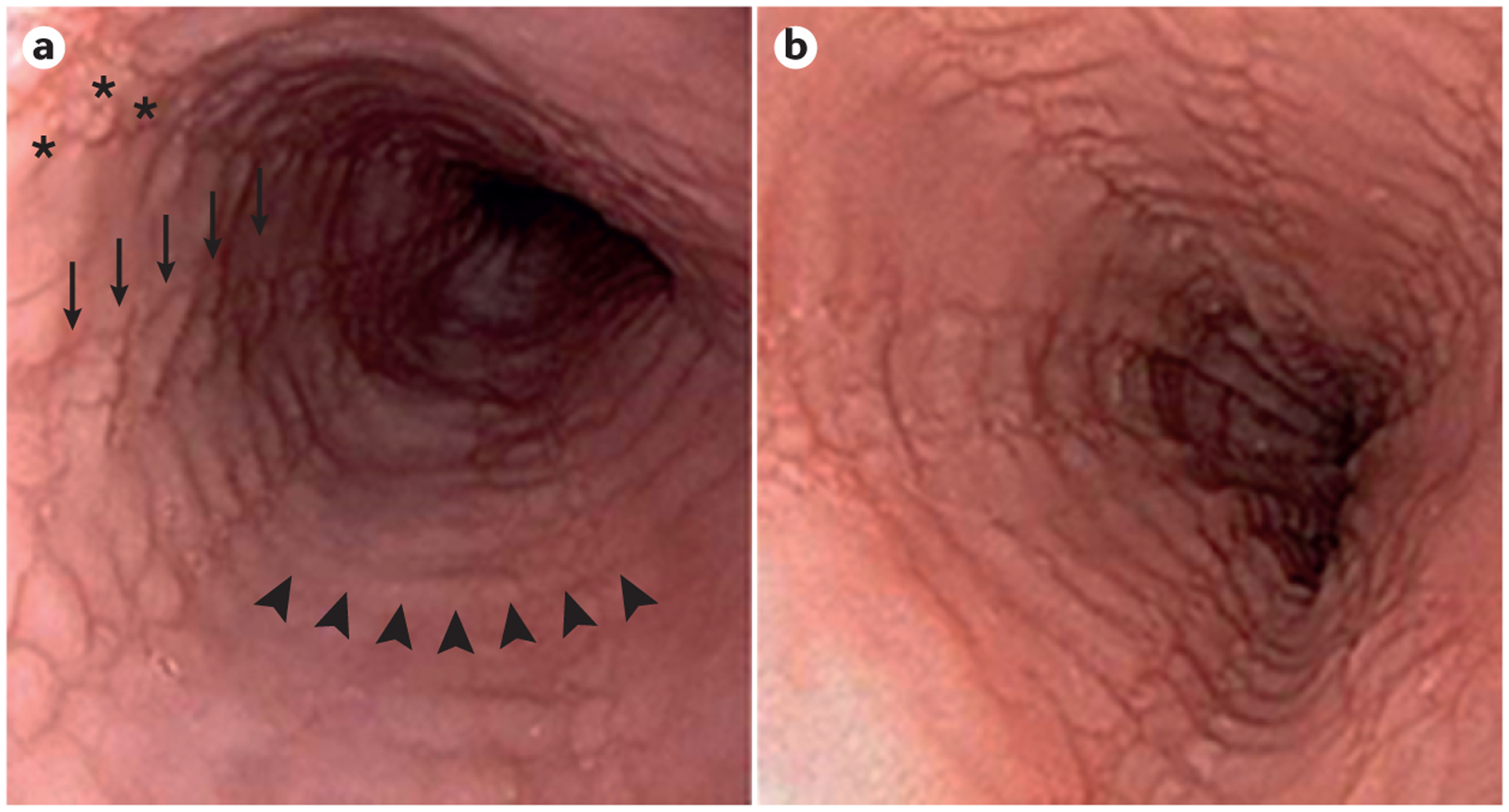
Paired endoscopic images of the oesophagus of a patient with eosinophilic oesophagitis (EoE) at diagnosis (part a) and after treatment with oral viscous budesonide at a dose of 1 mg twice daily (part b). At both time points, the patient remained symptomatic with dysphagia and there is no change in either the endoscopic findings (oedema (a diffuse loss of vascular markings), rings (arrowheads), subtle exudates (asterisks) and furrows (arrows) are seen at both time points) or histological findings (60 eos/hpf at diagnosis and 55 eos/hpf post-treatment). Eos/hpf, eosinophils per high-power field.