
Fig 4. PFFs injected in the OB induced an amplification and spread of pathology to interconnected regions, including the PCX.
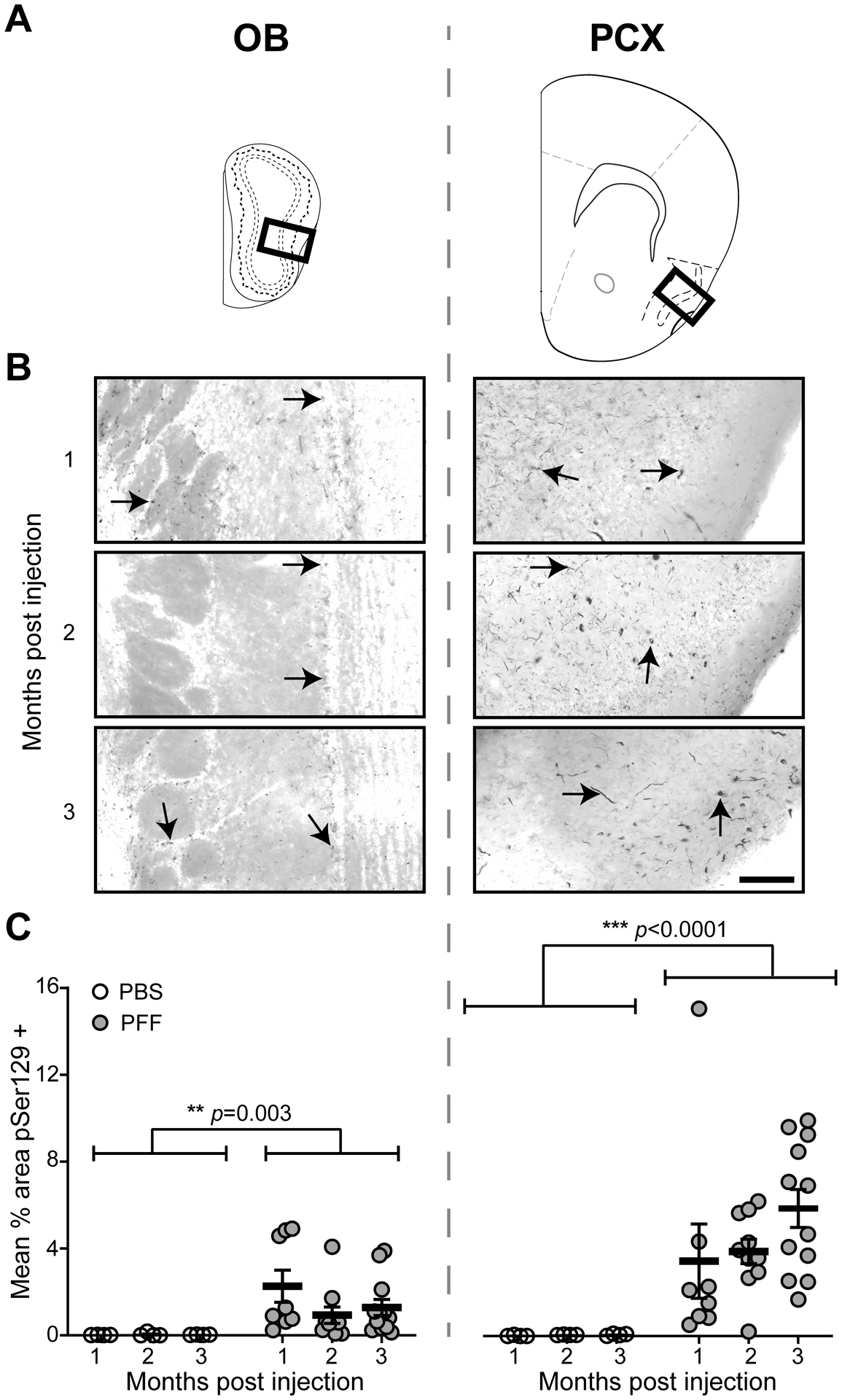
Pser129 immunofluorescence was used as an assay to detect pathological α-Syn. A, Coronal panel showing the regions (bold boxes) used for quantifying Pser129 expression level in the OB and PCX. B, Representative images of Pser129 immunofluorescence staining of the OB and PCX of mice that survived for 1, 2, or 3 months post PFF seeding. Arrowheads in the OB and PCX panels indicate areas of neuritic pathology. The images were gray-scaled and inverted to show pathology more readily, for illustration purposes of this figure only. C, Quantification of mean % area in the OB and PCX, showing Pser129 immunofluorescence in PFF and a subset of PBS injected mice that survived for 1 (PFF n= 8, PBS n= 4), 2 (PFF n= 10, PBS n= 4), and 3 (PFF n= 12, PBS n= 4) months post injection. Animals injected with PFFs had a significantly greater Pser129 immunopositive signal than the PBS injected animals, including in both the OB and PCX. Significant increase in mean % area Pser129 was observed in the PCX when compared to the OB. ***p ≤ 0.001 ANOVA followed by Tukey’s multiple comparison’s test.